
Chest X-Ray
Chest X-Ray
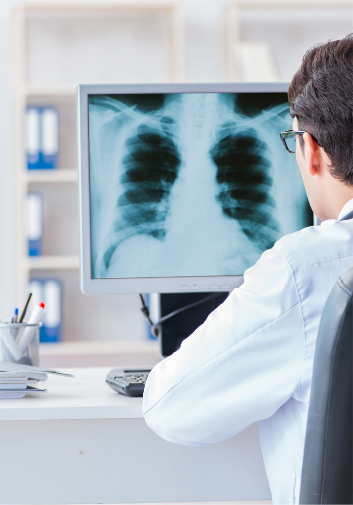
Chest X-ray is a non-invasive examination performed using special equipment in radiodiagnostic laboratories. X-rays (a type of electromagnetic radiation) are used to penetrate the human body (in the area of interest) and capture its “shadow” on special films (nowadays digital files) – this is possible as each body structure absorbs X-rays in a non uniform way.
The thoracic cage, the lungs, the heart and the large vessels are depicted in a two-dimensional fashion, Chest X-ray provides us information about the size of the heart, the presence of fluid in the lungs or the pericardium, the presence of abnormalities from the lungs, etc. A cardiologist may order this exam in the context of the differential diagnosis of shortness of breath, cough or fever. The findings are co-evaluated with the rest of the clinical-laboratory examination (physical examination, electrocardiogram, echocardiogram, etc.).
It is important to inform your doctor about the possibility of pregnancy prior to conducting any x-ray examination.