ECHOCARDIOGRAM
ECHOCARDIOGRAPHY(HeartTriplex)
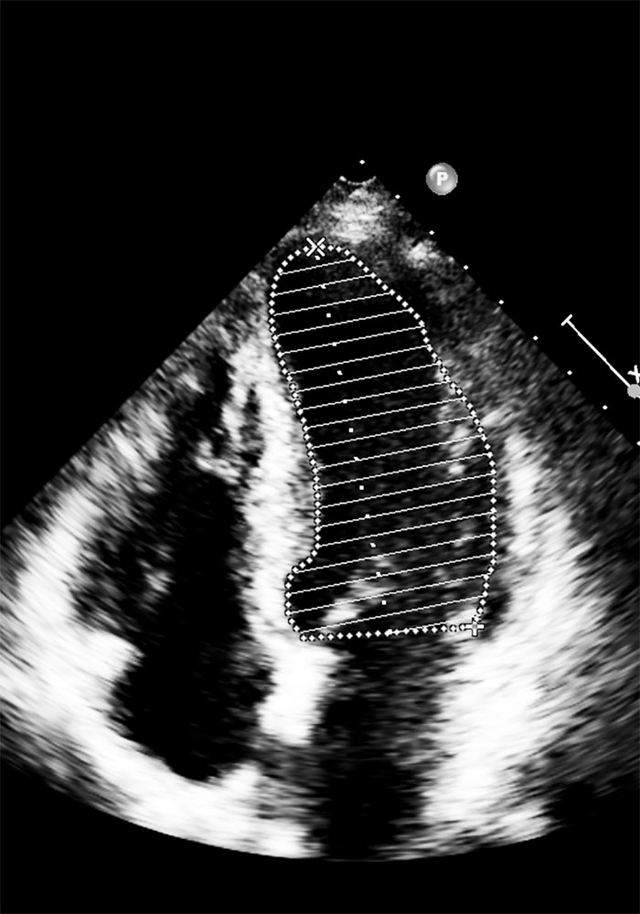
Cardiac Ultrasound (Echocardiogram or Heart Triplex) is one of the most valuable diagnostic examinations regarding the cardiovascular system. Information about the anatomy and functionality of the myocardium (heart muscle), heart valves, pericardium and great vessels are derived via echocardiography. An ultrasound machine is required in order to perform an echo scan. Typically, such a device involves an echo transducer (probe) and a central computing unit along with a monitor. The role of transducer is to create, send ultrasound waves and receive its reflections. The central unit utilizing special softare has the ablitiy to produce real-time images of heart structures and blood flow providing the physician with detailed information.
Transthoracic echocardiography is a non-invasive procedure for which no special preparation is required. The cardiologist asks the examinee to reveal his chest and lie on the examination bed turning to the left and raising his/her left hand (typically the patient is asked to lie down utilizing the left hand as a pillow). During the test electrodes may sometimes be placed in order to simultanesously monitor heart rhythm and frequency. Following, the cardiologist places the transducer in specific positions on the anterior and lateral chest wall of the examinee from which the heart (acoustic windows) may be visualised. A special gel is placed between the transducer and the chest, which is cleansed at the end of the examination.
Echocardiography is used to diagnose acute and chronic conditions, but also to monitor their course as long as potential response to treatment.
Transoesophageal Echocardiography
In some cases, the diagnostic capabilities of a transthoracic echocardiogram are not sufficient enough to reach a diagnosis; this is due the fact that a series of human body structures lie between the transducer and the heart. In such cases the attending cardiologist may request a more advanced imaging procedure such as a magnetic resonance imaging (MRI) of the heart or esophageal echocardiography. Transesophageal echocardiography follows the same technical methodology as transthoracic echocardiography. However, in the case of the transesophageal study, the transducer is advanced from the oral cavity to the esophagus and stomach (a process similar to gastroscopy), where they are close to the organ of interest, ie the heart. In other words, this is a mildly invasive test that requires proper preparation (the patient must be fasting for 6-8 hours prior to the examination) while mild sedation and/or anesthesia may be administered in more complex cases.