Written by Dimitrios Vrachatis, Specialist Cardiologist, PhD, National and Kapodistrian University of Athens.
What is an electrocardiogram?
An electrocardiogram (ECG) is the most commonly prescribed examination by cardiologists, often referred to simply as a cardiogram. A surface ECG records on paper the combined electrical activity of the heart.
How is an electrocardiogram performed?
A complete 12-lead surface electrocardiogram is recorded non-invasively in a procedure that takes only a few minutes.
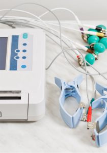
The person being examined is asked to uncover their chest, wrists and ankles. Special devices called electrodes are then placed at predefined points. The electrodes have small suction cups, clips for the arms and legs, or adhesive patches.
The electrocardiograph records the combined electrical signal produced by the heart cells during the different phases of the cardiac cycle.
Modern electrocardiographs use specialised equipment and software to filter out other electrical signals produced by the body, such as muscle activity, and interference, such as that caused by trembling. They may also provide an automated interpretation, which must always be confirmed by a cardiologist.
After recording, the electrocardiogram is printed, displayed on a screen or transferred to a computer.
Who needs an electrocardiogram, and when?
An electrocardiogram is an integral part of a cardiological examination. It is therefore performed on almost every visit to a cardiologist, whether for preventive assessment, such as a sports health certificate, or a scheduled review of a chronic condition. An ECG and its interpretation may also be requested before surgery; depending on the circumstances, a full cardiac assessment may be required.
In certain cases, such as suspected intermittent arrhythmias that occur for short periods, including atrial fibrillation, the cardiologist may recommend an ECG as soon as possible after symptoms begin, usually palpitations or an irregular pulse noticed when checking the pulse. A single ECG may not establish a diagnosis because it records only a short period. Longer recording with a 24-hour, 48-hour or longer Holter ECG may then be needed.
An ECG is also an urgent examination when acute symptoms occur, such as chest pain, shortness of breath, cold sweats, dizziness or fainting. In these circumstances, assessment also includes blood tests, an echocardiogram and further investigations.
Should I have an electrocardiogram or an echocardiogram first?
The two examinations are different. An electrocardiogram shows the electrical activity of the heart. An echocardiogram allows the heart to be seen and assessed on a screen.
They are complementary examinations that provide different types of essential information.
The cardiologist decides which examination is needed and in what order after taking the person’s medical history. The clinical assessment is an essential part of the examination process, beyond receiving an isolated test report.
How is an electrocardiogram interpreted?
Cardiologists evaluate ECG recordings systematically by recognising normal and abnormal patterns.
In simple terms, the cardiologist knows the expected shape of the familiar ECG line on the paper. They also relate the sequence of recorded electrical potentials to the functions and regions of the heart in order to draw the appropriate conclusions.
What information does an electrocardiogram provide?
The ECG provides indirect information about the integrity of the heart’s electrical system, including how electrical impulses are produced and conducted, the presence of acute myocardial ischaemia, the thickness of the heart walls and enlargement of the atria.
It is used in the diagnosis of an acute or previous myocardial infarction, pericarditis, myocarditis, arrhythmias, atrioventricular block, electrolyte disorders and toxic effects of medication, among other conditions.
What are the advantages of an electrocardiogram?
It is a non-invasive, simple, painless and quick examination. In Greece, its cost is also particularly affordable compared with other countries.
In addition to providing an overall picture, recording an arrhythmia on a complete 12-lead ECG is particularly important because it often establishes a definitive diagnosis and can guide specialised treatment.
An ECG also provides a valuable baseline for future comparison. Comparing an earlier recording with a new one, for example when symptoms develop, allows the findings and possible changes to be assessed with greater accuracy and diagnostic value.
What are the limitations of an electrocardiogram?
Although an ECG is one of the most valuable diagnostic tools in cardiology, it is a snapshot of the moment.
A normal ECG cannot rule out future adverse events or intermittent arrhythmias, which may occur only from time to time.
Limitations may also arise if the examination is not performed correctly: all leads may not have been recorded clearly, the settings may not provide all the information needed for a diagnostic recording, or the date and the person’s full name may not have been noted.
Is any preparation needed for an electrocardiogram?
No special preparation is required for an ECG. In exceptional cases, increased chest hair may require a little water or antiseptic, or local hair removal, to keep the electrodes stable. If needed, this is done on site with a disposable safety razor.
Are there any risks associated with an electrocardiogram?
An ECG is a non-invasive, painless recording procedure that poses no risk to the person being examined. The electrocardiograph only records the electrical activity produced by the heart; it does not deliver electricity to the heart.