
Written by Dimitrios Vrachatis, Specialist Cardiologist, PhD, National and Kapodistrian University of Athens.
What is coronary angiography?
Coronary angiography uses a contrast agent to show the lumen of the coronary arteries, the vessels supplying the heart muscle. It checks for narrowing, blockages and anatomical variations.
When is it needed?
- Urgently, for an acute myocardial infarction or unstable angina.
- As a planned investigation for coronary artery disease, for example in someone with chest pain and an abnormal stress echocardiogram, perfusion scan or exercise test, or newly diagnosed heart failure.
- As a planned preoperative assessment before valve surgery.
How is it performed?
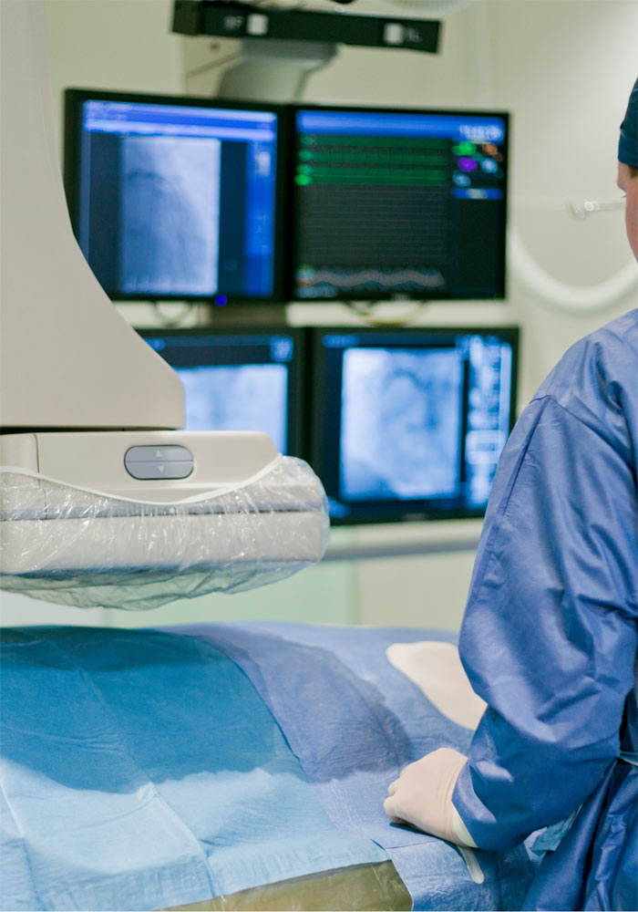
In the catheterisation laboratory, you remove your clothes and lie on the procedure table. Preparation includes shaving, antiseptic cleaning and insertion of an intravenous cannula if not already done. Vital signs are closely monitored throughout.
The interventional cardiologist advances a diagnostic catheter to the aortic root, where the coronary arteries originate, to inject contrast.
A peripheral artery is punctured under local anaesthesia and a special atraumatic guidewire is advanced. A flexible diagnostic catheter, approximately 2 mm in diameter, passes over the wire, which is then removed.
The catheter is positioned at the openings of the left and right coronary arteries to perform the examination.
How are the arteries visualised?
Once access is established:
- Contrast is injected into the coronary arteries while
- Brief fluoroscopy is performed, usually for 2–3 heartbeats.
This records a moving two-dimensional image, like a short video. The process is repeated from different, perpendicular projections so that the cardiologist can assess the vessel lumen.
Can other techniques be used?
Additional imaging, such as intravascular ultrasound (IVUS), or functional assessment, such as fractional flow reserve (FFR), can be performed during the same session.
What if I need angioplasty or a stent?
If angiography shows a need for angioplasty and stenting, this can often be performed in the same session. Complex anatomy or other considerations may require it to be scheduled separately.
Is access through the wrist or the groin?
Most centres now use the radial artery near the right wrist.
If the right arm presents anatomical difficulties, the left can be used. Left-sided access is also preferred in patients with previous bypass surgery.
If wrist access is not possible, the femoral artery in the right or left groin is used.
What are the advantages of each approach?
When successfully completed, the diagnostic value is the same.
The main difference is earlier mobilisation after wrist access. With the radial approach, patients can get up within 2–3 hours and may go home the same day if no angioplasty was performed.
Femoral access generally requires lying still for at least 7–8 hours to achieve haemostasis, with weights applied over the puncture site. Femoral puncture also carries a higher complication risk than radial puncture.
How long does it take?
The main examination takes no more than 15–20 minutes. Additional time is needed for preparation and transfer because the catheterisation laboratory follows operating-theatre standards of antisepsis and safety.
Is it painful?
The procedure is generally painless under local anaesthesia. The anaesthetic injection may briefly sting. You may feel local pressure as the sheath, the temporary access tube, is inserted despite the anaesthesia.
Report any chest pain or breathlessness during or after the examination immediately to the medical or nursing team.
After femoral access, the doctor applies local pressure to stop bleeding, which can be uncomfortable. After radial access, a special wristband applies gentle pressure without significant pain.
When are the results available?
Images are recorded digitally, for example on a CD or flash drive, rather than the film used previously. They are available immediately and should always be given to the patient. They are useful for a second opinion and as part of the medical history, even years later. The interventional cardiologist prepares the written report immediately or subsequently.
What is a catheterisation laboratory?
A specially equipped hospital facility staffed by trained nurses and technologists, where coronary angiography is performed.
What preparation is needed?
- Do not eat during the hours preceding the examination.
- Good hydration in the preceding and following days helps reduce contrast-related kidney injury. Some patients receive intravenous hydration in hospital.
- Tell your cardiologist beforehand about any known or suspected contrast allergy so that appropriate medication can be arranged.
- Tell your cardiologist if you take anticoagulants; you will receive specific instructions.
- For a planned examination, shaving the groin beforehand allows femoral access if the radial approach is not feasible.
What are the risks?
As with any invasive investigation, risks exist but are outweighed by the expected benefit. They include:
- Bleeding or haematoma at the arterial puncture site
- Complications while advancing equipment to the aorta and coronary arteries, such as dissection, vessel injury or stroke
- Contrast-related kidney toxicity or allergic reactions
The procedure also involves ionising radiation.
To indicate their approximate scale, risks for a stand-alone diagnostic study include:
- Less than 0.1%, or one in 1,000, for a procedure-related acute coronary event
- 0.05–0.1% for stroke
- Less than 0.05%, or five in 10,000, for death
When angiography is clearly indicated, these risks are lower than the risks of omitting the examination.