
Written by Dimitrios Vrachatis, Specialist Cardiologist, PhD, National and Kapodistrian University of Athens.
What is mitral regurgitation?
Mitral regurgitation is abnormal backward flow of blood from the left ventricle into the left atrium through the mitral valve.
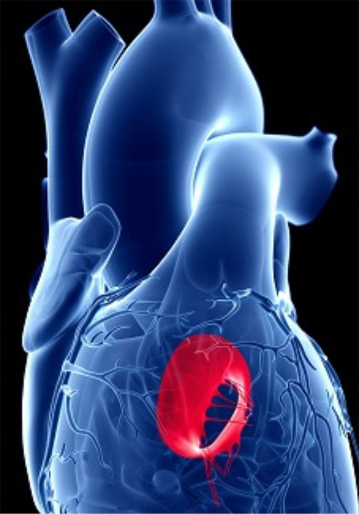
What is the mitral valve?
The mitral valve connects the left atrium and left ventricle. Its complex structure, known as the mitral apparatus, includes two leaflets, papillary muscles and chordae tendineae.
Normally, after blood passes from the atrium into the ventricle during relaxation, the valve closes tightly to prevent backward flow.
How is mitral regurgitation diagnosed?
Diagnosis uses an echocardiogram. An electrocardiogram or prolonged Holter monitoring may also be performed if arrhythmias are suspected.
Advanced cases may require transoesophageal echocardiography or cardiac MRI to establish severity and mechanism accurately.
What are the symptoms?
Mitral regurgitation can be misleading: it may remain asymptomatic for a long time and only be diagnosed after heart failure develops.
Acute, sudden regurgitation is very different from chronic regurgitation, which develops gradually.
Chronic Mitral Regurgitation
When regurgitation develops gradually, the heart has time to adapt. The left atrium enlarges and the left ventricle develops eccentric hypertrophy.
This maintains adequate function for a time, until compensation eventually fails. Symptoms develop gradually and may include:
- Palpitations due to extra beats or atrial fibrillation
- Fatigue or breathlessness
- Chest pain
- Dizziness or feeling faint
- Swollen legs
- Needing more than one pillow to sleep
Acute Mitral Regurgitation
When regurgitation develops suddenly, the left atrium cannot accommodate the increased blood volume. Pressure rises and is transmitted to the lungs, causing acute pulmonary oedema, or acute heart failure. Emergency surgery is therefore required.
Classification
By cause, regurgitation is:
- Primary: damage to a component of the valve apparatus
- Secondary: caused by left ventricular dysfunction
By severity, it is classified as:
- Mild
- Moderate
- Severe
Causes
This valve disease has several possible causes:
- Congenital valve abnormalities
- Mitral valve prolapse
- A myocardial infarction involving the papillary muscles
- Rupture of the chordae tendineae
- Rheumatic fever
- Infective endocarditis
- Cardiomyopathies, among others
Follow-up
Once moderate mitral regurgitation is diagnosed, regular follow-up is needed to identify deterioration and any need for intervention promptly.
What treatment is available?
Treatment is complex. Intervention, such as valve repair or replacement, depends on the cause and the stage of cardiac enlargement and functional impairment. It concerns severe regurgitation only. Medication is not specific to regurgitation itself: it may treat the underlying cause or associated heart failure.
Surgical Repair or Replacement?
Repair is preferred over prosthetic replacement when technically feasible.
Choosing the right time is critical: intervention should take place as soon as symptoms or signs of functional or structural deterioration appear.
This applies to chronic regurgitation. Acute regurgitation requires immediate cardiac surgery.
Mechanical or Biological Prosthetic Valve?
The choice depends on age and individual characteristics. Mechanical valves last longer, reducing the likelihood of another operation, but require lifelong anticoagulation with acenocoumarol or warfarin. Biological valves do not require anticoagulation for the valve itself but have a shorter lifespan.
Are minimally invasive options available?
For some patients with symptomatic severe regurgitation who are at high surgical risk, catheter-based techniques such as edge-to-edge repair using devices like MitraClip may be possible.
Mitral Regurgitation and Dental Treatment
The cited European Society of Cardiology guidelines (ESC 2015) do not recommend preventive antibiotics for patients with mitral regurgitation. Good oral hygiene is nevertheless important.
Mitral Regurgitation and Exercise
Sports participation, provided there are no valve-related symptoms, is described as follows in the cited ESC 2021 guidelines:
- Mild: unrestricted participation.
- Moderate: unrestricted participation may be discussed if specific criteria for left ventricular function and structure are met on echocardiography and an exercise test shows a normal response.
- Severe: participation in low- or moderate-intensity sports only may be discussed if the same criteria for ventricular function and structure are met on echocardiography and an exercise test is normal.