
Written by Dimitrios Vrachatis, Specialist Cardiologist, PhD, National and Kapodistrian University of Athens.
What is an echocardiogram?
An echocardiogram is one of the most valuable cardiovascular examinations. It displays the heart on a specialised computer screen. Without further qualification, the term usually means a resting transthoracic study, performed from outside the body: a painless, non-invasive procedure.
This differs from stress echocardiography, which examines the heart during medication-induced or physical stress.
In specific cases, a transoesophageal study may be needed. Its procedure resembles gastroscopy; more information is provided below.
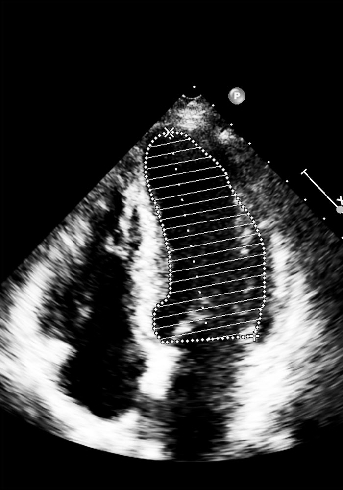
What does an echocardiogram show?
It provides information about:
- Cardiac anatomy, including wall thickness and dimensions
- Movement of individual heart walls
- Overall myocardial function, particularly left ventricular ejection fraction
- Blood clots within the chambers
- Valve structure and integrity
- Valve function
- Abnormal communications between heart chambers
- The pericardium, including any fluid around the heart
- Major vessels: ascending aorta, aortic arch and pulmonary artery
What is it used for?
It diagnoses acute and chronic conditions, including:
- Heart failure
- Coronary artery disease
- Valve disease
- Cardiomyopathies
- Pericarditis
- Myocarditis
- Ascending aortic aneurysm
It also monitors their course and response to treatment, and follows the effects on the heart of conditions such as hypertension or atrial fibrillation.
How is it performed?
The equipment includes a probe, a central signal-processing unit and a screen.
The probe, resembling a large marker pen, is placed against the chest. It emits harmless ultrasound waves at 2.5–5 MHz, inaudible to humans, and records returning echoes.
The cardiologist asks you to remove clothing from the waist up, lie on your left side and raise your left arm, often resting your head on it as though it were a pillow.
Electrodes may be attached to monitor heart rate and rhythm using a single-lead ECG.
The probe is placed at specific positions on the front and side of the chest, known as acoustic windows. Gel between the probe and skin helps the examination and is wiped off afterwards.
The computer analyses the returning signals in real time and uses specialised algorithms to display the heart and blood flow, providing detailed information to the examiner.
Is preparation needed?
No special preparation is required for transthoracic echocardiography. It is generally painless, although pressure from the probe may occasionally cause minor, temporary discomfort.
When will I receive the results?
The examination takes approximately 10–30 minutes. By the end, the cardiologist usually understands the findings and can explain them verbally. Complex cases may require additional measurements on the stored images and videos.
A full written report takes time to prepare but is usually provided immediately afterwards. A complete report, rather than brief notes, is important as a record for future comparisons.
Who needs an echocardiogram, and how often?
For healthy adults, a one-time examination can provide a useful baseline.
People with established cardiac conditions may need repeated studies. The interval, whether 3 or 6 months, one or two years or longer, depends on the condition and when it was diagnosed.
Intervals are usually shorter after a new diagnosis, such as aortic regurgitation or aortic stenosis, and may become longer later. More frequent reassessment is also needed when approaching thresholds for treatment decisions, for example in valve disease.
A new symptom or deterioration of a previously stable condition usually requires another echocardiogram.
Are there risks?
A resting transthoracic echocardiogram is painless, non-invasive and uses no ionising radiation. It carries no associated risk and is safe even during pregnancy or in children.
What are its limitations?
Like an electrocardiogram, a resting echocardiogram is a snapshot of a moment already in the past. It cannot exclude future events.
If damage is not present at the time of examination, or lies outside what the test assesses, it cannot be identified.
For example, someone with significant coronary disease may have a completely normal resting study if the heart receives enough oxygen at rest. This does not reduce the examination’s value, but both patient and cardiologist should understand its capabilities. Conversely, early recognition of a subtle finding, such as reduced movement of a wall, can allow timely treatment and prevent a worse outcome.
After taking a detailed history, the cardiologist may recommend additional non-invasive tests, such as an exercise test, stress echocardiogram or myocardial perfusion scan, or invasive tests such as coronary angiography.
Why is it sometimes called a cardiac “triplex”?
The term reflects the technologies combined in echocardiography: two-dimensional black-and-white imaging, pulsed or continuous-wave Doppler, and colour flow imaging. Red and blue indicate opposite directions of flow relative to the probe, while colour intensity represents velocity. “Triplex” is a historical term that remains common in Greek everyday usage, alongside “heart echo”, instead of echocardiogram.
What is transoesophageal echocardiography?
Sometimes a transthoracic study cannot provide a diagnosis because body structures lie between the probe and the heart.
The cardiologist may request another imaging method, such as cardiac MRI or transoesophageal echocardiography.
The ultrasound principle is the same, but a specialised probe passes through the mouth into the oesophagus and stomach, similarly to gastroscopy. These structures are very close to the heart.
This is a mildly invasive examination requiring preparation, including fasting for 6–8 hours beforehand. Mild sedation or, in more difficult cases, anaesthesia may be used.