
Written by Dimitrios Vrachatis, Specialist Cardiologist, PhD, National and Kapodistrian University of Athens.
What is CT coronary angiography?
Cardiac computed tomography, or CT coronary angiography, is a non-invasive method of imaging the coronary arteries to assess narrowings that cause ischaemia.
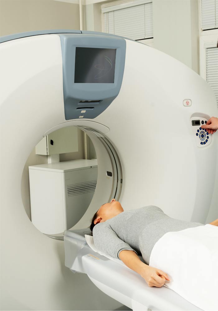
How is it performed?
A peripheral intravenous catheter is inserted to administer contrast medium while a CT scan focused on the heart is performed.
Who is it for?
CT coronary angiography is mainly indicated for people with a low to moderate clinical suspicion of coronary artery disease. This is assessed from symptoms — such as chest pain, shortness of breath or fainting — and conventional risk factors, including hypertension, dyslipidaemia, diabetes, smoking, obesity and family history.
It is also useful when complex anatomy is likely, for example after bypass surgery, where both patent and occluded grafts can be seen, or when an abnormal course of the native coronary arteries is suspected.
CT angiography of the aorta and major arteries, performed using a different scanning protocol, may form part of the pre-procedure assessment for transcatheter aortic valve implantation (TAVI).
What are the advantages?
The main advantage is its non-invasive nature. Only a peripheral venous catheter is needed, and potential risks relate to the contrast medium and ionising radiation. Structures outside the heart are also imaged.
The examination has a high negative predictive value: a negative result is particularly reliable and helps rule out coronary artery disease.
What are the disadvantages?
Disadvantages include:
- Radiation exposure.
- Patient-related technical difficulties that may make a study non-diagnostic, such as a high calcium burden, obesity, a rapid heart rate or arrhythmias. Arrhythmias are less problematic with high-resolution multislice CT scanners.
- The need for conventional invasive coronary angiography if CT findings suggest significant coronary artery disease.
Where is it performed, and how long does it take?
It is performed in a CT scanner. The scan itself takes less than half a minute. Insertion of the intravenous catheter and taking a medical history take place beforehand.
Is preparation needed?
The acceptable upper heart rate depends on the scanner’s capabilities. A rapid heart rate is undesirable, so a small dose of heart-rate-lowering medication may be needed in consultation with the treating doctor and the imaging centre.
Avoid heavy meals and stimulant drinks, such as coffee and tea, for 3–4 hours beforehand.
The possibility of pregnancy must always be checked because the examination involves radiation.
When can I return to my activities?
No hospital stay is needed. You can return to your activities the same day.
What should I be aware of?
If kidney disease is present, adequate hydration is important because contrast medium is administered.
When will I receive the results?
The CT scan records images of the heart, but specialised processing and reconstruction on dedicated computers are required before the study can be assessed and the report issued.