ELECTROCARDIOGRAM

ELECTROCARDIOGRAM (ECG)
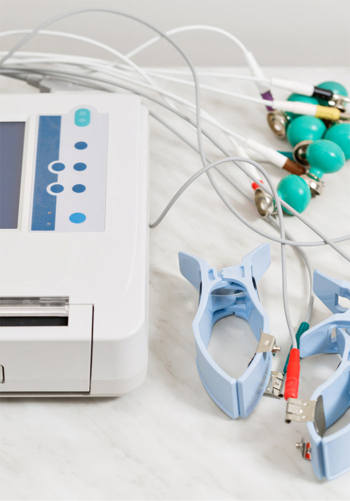
Electrocardiogram (ECG) is the most commonly prescribed examination by cardiologists. ECG is recorder non-invasively in a procedure which requires a few minutes to be completed. The examined person will be asked to undress so that his/her chest is revealed as well as regions around the hands and the ankles. Following special devices, called electrodes, are placed in predefined spots. Electrodes are equiped with special suction cups (or calipers for the upper and lower extremities) or special patches.
Following, the electrocardiograph is set to record the component of the electrical signal induced during the various phases of the cardiac cycle. Modern electrocardiographs are designed and assisted by special software in order to exclude other electrical signals that may be produced by the human body (e.g. potentials from muscles cells) or various artifacts. Following recording, the electrocardiogram is printed or shown in a screen or transferred in a PC.
Cardiologists evaluate ECG recordings under a systematic approach based on patterns’ recognition. The ECG provides us with indirect information about the integrity of the electrical heart system -regarding both production and/or conduction-, the presence of acute myocardial ischemia, the thickness of the myocardial walls, etc. The recording of possible arrhythmias on a 12-lead electrocardiogram is of particular importance because it definitively sets the diagnosis and guides treatment.
However, one shall keep in mind that an ECG is just a picture of the moment. It is undoubtedtly, one of the most valuable diagnostic tools in cardiology, but a normal ECG by no means can foresee future adverse events neither preclude intermittent arrhythmias – i.e. arrhythmias that are not constantly present.
No special preparation is required to perform an ECG, although increased chest hair growth in men may sometimes necessitate the use of a small amounts of water or -in extreme cases- local hair removal to keep the electrodes stable.
