
COMPUTED TOMOGRAPHY CORONARY ANGIOGRAPHY
CARDIAC COMPUTED TOMOGRAPHY
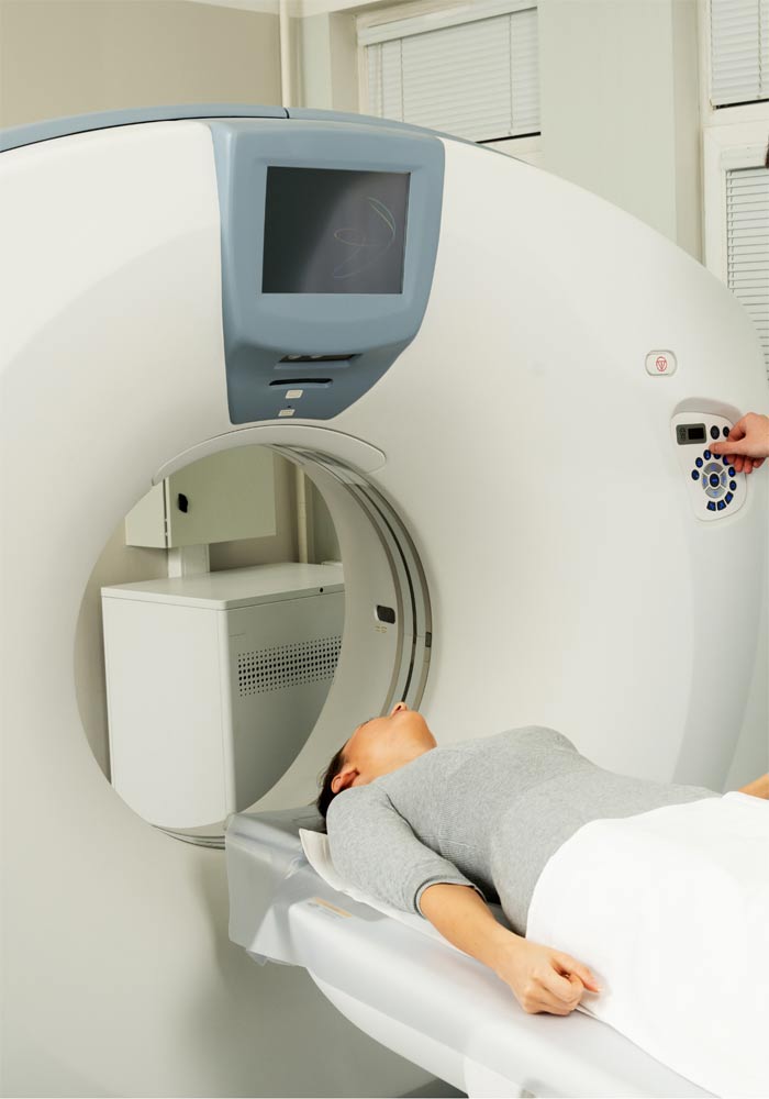
Computed tomography (CT) of the heart is a non-invasive method fot imaging utilized in the context of coronary artery disease evaluation. To perform a cardiac CT, a peripheral venous catheter is placed via which contrast agent is administered. Currently it is mainly indicated for the investigation of people with low to moderate clinical suspicion of coronary heart disease.
The main advantage of the method is its non-invasive nature. Disadvantages of the method include the exposure to radiation along with some technical issues (which gradually decrease with technological progress) but also the need to perform “traditional” invasive coronary angiography in the case that findings compatible with significant coronary heart disease are revealed.
It is performed on a CT scanner and the examination (scan) requires less than 30 seconds. The examination is preceded by the placement of the catheter and medical history taking. Patient returns to his daily routine on the same day. In the case of concomitant kidney disease, adequate hydration is important due to contrast media administration.
CT angiography of the aorta and large arteries of the human body, which is performed with a different scan protocol, may be performed as part of the preoperative evaluation of patients who are to undergo transcatheter aortic valve implantation (TAVI) or other structural heart disease procedures.