
CORONARY ARTERY ANGIOGRAPHY
CORONARY ARTERY ANGIOGRAPHY – LEFT HEART CATHETERIZATION
Coronary angiography (or coronary arteriography) refers to the imaging of the lumen of the arteries that provided oxygenated blood to the heart muscle, that is, the coronary arteries. For this scope a special substance called a contrast agent, is intraarterially administered. The purpose of the imaging of the lumen of the coronary arteries is to check for stenoses, obstructions or anatomical variations.
The contrast media is injected into the coronary arteries and at the same time radioscopy is performed (usually with a duration equivalent to the time needed for 2-3 heart contractions). Thus, a continuous two-dimensional image recording (like a small video) is produced. This procedure is repeated from different (vertical to each other) projections so that the invasive cardiologist draws the appropriate conclusions about the lumen of the vessels.
The invasive cardiologist in order to “approach” the coronary arteries and perform the required contrast injections (and / or to apply complementary novel imaging techniques such as intracoronary echocardiography – IVUS – or fractional flow reserve -FFR-) should first advance to the aortic root a special diagnostic catheter; coronary arteries rise from the aortic root. Access to the central arterial system is succeded by a puncture of a peripheral artery (under local anesthesia) and advancement of a non-traumatic angiography wire, over which the diagnostic catheter is subsequently advanced. Finally the the coronary wire is withdrawn. Then, the diagnostic catheter with special manipulations is placed at the ostium of the left and right coronary artery and the examination is performed. If this is indicated, percutaneous coronary intervention (angioplasty – stent placement) can be performed in the same session.
Nowadays, in most centers the peripheral artery selected for vascular access is an artery in the right upper limb near the wrist called radial artery. In the case of anatomical difficulties the option to gain vascular access via the contralateral upper extremity or via the right or left femoral artery may be opted for.
Coronary angiography may also be performed on an emergency basis (e.g. in acute myocardial infarction) or on a scheduled basis (e.g. in the context of investigating the presence of coronary artery disease or prior to cardiac surgery).
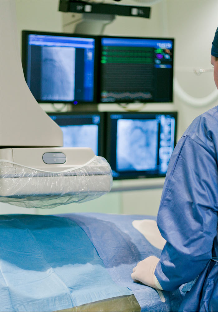
The procedure is conducted in an area specially equipped called catheterization laboratory and is most of the cases located whithin a hospital. The result of the test is now recorded on digital media – in the past it was recorded on special films.
Like any invasive examination, it is accompanied by risks which, however, are overcome by the expected benefit. These risks may be associated with access point complications (bleeding and / or hematoma), advancement of wires/catheters to the aorta and the coronary arteries (dissection, vascular rupture, stroke), and with nephrotoxicity due to the contrast medium or an allergic reaction to it. It should be underlined that this test is accompanied by the utilization of ionizing radiation. To understand the order of magnitude, the risk is less than 0.1% for acute iatrogenic myocardial infarction, 0.05 – 0.1% for stroke or less than 0.05% death in the case of a diagnostic coronary angiography. As already noted, the risk for complications is lower than the risk of not conducting the coronary angiography (and hence missing an potentially important diagnosis), when it’s indication is well documented.